
Written by a Caring Mom at Infant Pamper
At seven weeks, pregnancy can start to feel more real. Some symptoms might feel stronger. You may find yourself opening a pregnancy app again and again. You may also wonder what is going on inside your body right now.
A 7 Weeks pregnant ultrasound can give an early view of how things are going. It may help estimate how far along you are. It can also show where the pregnancy is located. In some cases, early signs of a heartbeat may be seen.
It is also normal to feel worried before the appointment. This is true even more so if it is your first pregnancy, or if you have been waiting a long time for the scan. Seven weeks is still early, so the scan may seem basic. Even then, it can still show key early milestones in pregnancy.
Table of Contents
- Why Might You Have an Ultrasound at 7 Weeks?
- What Can a 7 Weeks Pregnant Ultrasound Show?
- How Big Is Baby at 7 Weeks?
- What If You Thought You Were 7 Weeks but Measure Earlier?
- Abdominal vs. Transvaginal Ultrasound at 7 Weeks
- What If Very Little Is Visible?
- When Should You Contact Your Healthcare Provider?
- Frequently Asked Questions
- Final Thoughts
- Medical Disclaimer
Why Might You Have an Ultrasound at 7 Weeks?
Not every pregnancy includes an ultrasound at exactly seven weeks.
Routine scan timing varies depending on where you live and your healthcare provider. An early scan may be offered because your doctor wants a closer look at how the pregnancy is progressing.
You may have an early ultrasound if:
- your dates are uncertain
- you have irregular menstrual cycles
- you have bleeding or pelvic pain
- you became pregnant through IVF or fertility treatment
- you have experienced a previous pregnancy loss
- you have had an ectopic pregnancy before
- your doctor wants to confirm the pregnancy is inside the uterus
- twins or multiples are suspected
Sometimes there is no worrying reason at all. Your healthcare provider may simply want to confirm your gestational age.
What Can a 7 Weeks Pregnant Ultrasound Show?
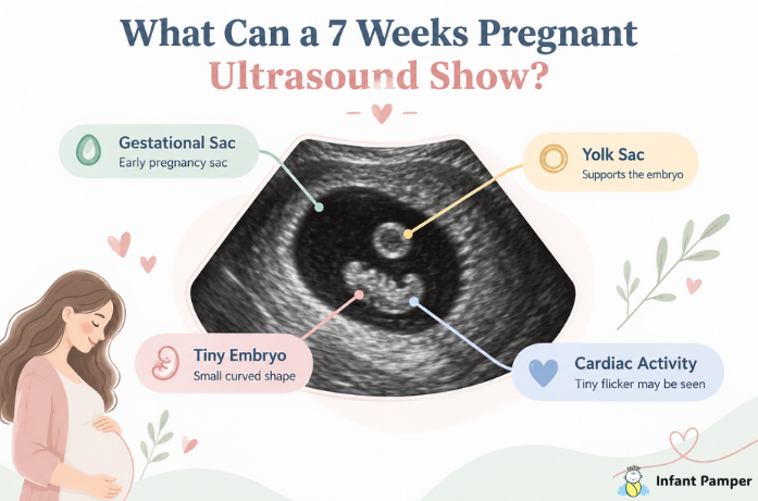
A seven-week ultrasound can show several important early pregnancy structures, although the exact picture can vary depending on your true gestational age and whether the scan is abdominal or transvaginal. At this stage, your baby is still very small, so the image may look more like a tiny curved shape than a recognizable baby.
Gestational Sac
The gestational sac is usually one of the first structures seen in early pregnancy. It appears as a dark, round or oval area inside the uterus.
Seeing the sac in the uterus helps your healthcare provider confirm that the pregnancy is developing in the expected place. They may also look at its size and shape as part of the overall assessment.
Yolk Sac
The yolk sac is a small round structure found inside the gestational sac. It helps support the embryo during the earliest weeks before the placenta is fully taking over that role.
On the ultrasound, it may look like a tiny bright ring. Its presence can give your healthcare provider another useful sign that the early pregnancy is developing.
Tiny Embryo
By around seven weeks, the embryo may be visible next to the yolk sac. It can look like a small bean, curved line, or little shape tucked inside the gestational sac.
The sonographer may measure the embryo from top to bottom using the crown-rump length, or CRL, which can help estimate how far along the pregnancy is.
Cardiac Activity
Early cardiac activity may also be visible at this stage, especially during a transvaginal ultrasound. It often appears as a tiny flicker within the embryo.
You may see this movement without hearing any sound, and that can still be completely normal. Your sonographer may simply measure the activity on the screen rather than play an audible heartbeat.
According to NCBI Bookshelf (StatPearls), the gestational sac is typically the first sign seen on ultrasound, followed by the yolk sac and fetal pole, with cardiac activity usually becoming visible by around the sixth or seventh week.
How Big Is Baby at 7 Weeks?
At seven weeks, your baby is still very small.
The NHS compares a seven-week embryo to roughly the size of a grape.
Even though your baby is tiny, important development is already happening.
Early structures that will eventually become the brain, face, arms, legs, and organs are beginning to form.
Most of that development is far too small to see clearly on your ultrasound.
This early scan is mainly about confirming pregnancy location, gestational age, and early development rather than checking detailed anatomy.
What Do Common Ultrasound Terms Mean?
Ultrasound reports can sometimes feel like they are written in another language.
Here are a few common words you may hear.
Gestational sac: The fluid-filled space surrounding the developing pregnancy.
Yolk sac: A small structure that helps support the embryo during early pregnancy.
Embryo or fetal pole: The developing baby seen beside the yolk sac.
CRL: Crown-rump length, a measurement from the top of the embryo to its lower end.
Gestational age: Your estimated number of weeks and days pregnant.
Cardiac activity: The early movement associated with the developing heart.
Knowing these terms beforehand may help you feel more confident during the appointment.
Why Does Crown-Rump Length Matter?
During your ultrasound, the sonographer may take a tiny measurement from the top of your baby’s head area to the bottom of the body. This is called the crown-rump length, or CRL.
It may sound like just another medical term, but CRL can help your healthcare provider estimate how many weeks pregnant you are. This is especially useful if your cycles are irregular or you are not completely sure when your last period started.
And if the measurement shows you are a few days earlier or later than you expected, try not to worry straight away. Small differences in early pregnancy dates are quite common.
What If You Thought You Were 7 Weeks but Measure Earlier?
This is one of the most common reasons parents become worried after an early ultrasound.
You may arrive expecting to measure exactly seven weeks and then be told the baby looks closer to six weeks and four days.
Pregnancy dating is not always exact.
Weeks of pregnancy are normally counted from the first day of your last menstrual period. The American Pregnancy Association also notes that pregnancy dating begins from the first day of your last menstrual period rather than the day conception actually occurred. That calculation assumes ovulation happened around a typical time.
But bodies do not always follow a perfect schedule.
You may have:
- ovulated later than expected
- had a longer menstrual cycle
- had an irregular cycle
- remembered your last period slightly incorrectly
- conceived later than you assumed
Even a few days can make a noticeable difference during very early pregnancy.
Your healthcare provider will look at the complete picture, including measurements and follow-up scans when needed.
Abdominal vs. Transvaginal Ultrasound at 7 Weeks
You may expect your first ultrasound to be done across your stomach, but early pregnancy scans can also be performed vaginally.
Abdominal Ultrasound
For an abdominal ultrasound, gel is placed on your lower belly and the probe moves across the skin.
Some clinics ask you to arrive with a full bladder because it can improve the view.
Your clinic should tell you how to prepare.
Transvaginal Ultrasound
During a transvaginal ultrasound, a slim covered probe is gently inserted into the vagina.
It may sound uncomfortable if you have never had one before, but it is commonly used in early pregnancy because it gives a closer and clearer view of the uterus.
A transvaginal scan may make it easier to see the gestational sac, embryo, and early cardiac activity. Cleveland Clinic explains that early pregnancy ultrasounds around seven to eight weeks are often performed transvaginally and may be used to confirm pregnancy, check for multiples, measure the developing baby, and help estimate gestational age.
Tell the sonographer if you feel uncomfortable or need them to pause.
You are always allowed to ask questions about what is happening.
What If You See Cardiac Activity but Hear Nothing?
It is easy to expect that seeing cardiac activity also means you will hear a heartbeat, but that is not always the case this early.
At seven weeks, the sonographer may be able to see a tiny flicker on the screen and measure the activity without playing any sound. So if the room stays quiet, try not to worry.
If you are unsure, simply ask whether cardiac activity is visible and what the sonographer is able to measure.
What If Very Little Is Visible?
Sometimes an early ultrasound does not show as much as you expected, and that can feel worrying. But at this stage, even being a few days earlier than you thought can make a noticeable difference in what appears on the screen.
The view can also depend on the position of your uterus and embryo, as well as whether the scan is abdominal or transvaginal. If your healthcare provider needs a clearer picture, they may suggest another ultrasound after some time.
Waiting can be hard, but try not to compare your scan with images online. Every pregnancy has its own timing and measurements.
Can Twins Be Seen at 7 Weeks?
Yes, twins can sometimes be seen on an ultrasound at seven weeks. The sonographer may notice two gestational sacs, two embryos, or other early signs of a twin pregnancy.
If twins are suspected, they will look closely at the sacs and early pregnancy structures to understand how the pregnancy is developing. And if the scan just looks like a few tiny circles and shapes to you, that is completely normal early ultrasound images can be hard to understand without training.
How to Prepare for Your Scan
There is usually very little you need to do.
Your clinic may give you instructions about whether to arrive with a full bladder.
You can also make the appointment easier by bringing or knowing:
- the first day of your last menstrual period
- your usual cycle length
- details of previous pregnancies
- information about fertility treatment if relevant
- medications and supplements you take
- any symptoms you want to discuss
Wear comfortable clothing and keep a few questions saved on your phone.
Appointments can feel emotional, and it is surprisingly easy to forget what you wanted to ask.
Helpful Questions to Ask
You do not need a long list.
A few simple questions can help you leave feeling more informed.
You might ask:
- Is the pregnancy inside the uterus?
- How many weeks and days am I measuring?
- Can you see cardiac activity?
- What is the crown-rump length?
- Is there one baby or more than one?
- Do I need another ultrasound?
Do not feel embarrassed about asking for an explanation.
You are not expected to understand ultrasound images on your own.
When Should You Contact Your Healthcare Provider?
Mild cramping or spotting can happen during early pregnancy, but it is always okay to contact your healthcare provider if you are worried.
Seek urgent medical care for severe or worsening pelvic or abdominal pain, heavy bleeding, fainting, significant dizziness, shoulder-tip pain, or symptoms that feel serious or unusual.
Some of these symptoms can require urgent evaluation, including checking for ectopic pregnancy.
Trust yourself enough to ask for help when something does not feel right.
Frequently Asked Questions
Final Thoughts
A 7 weeks pregnant ultrasound can be a small appointment with a lot of emotion packed into it. Seeing a tiny embryo or that first flicker of cardiac activity may make the pregnancy feel real in a whole new way.
If your dates are a little different from what you expected, try not to compare your scan too closely with someone else’s. Early pregnancy can vary by several days, and your healthcare provider will look at the full picture rather than one number on the screen.
Use the appointment to ask questions, understand what was seen, and find out what comes next. Your baby is still very small, but this early stage is already full of important growth and change.
For more gentle, practical pregnancy tips and week-by-week guidance, explore more helpful articles on Infant Pamper.
Medical Disclaimer
Information on Infant Pamper is for general educational purposes only and should not replace medical advice, diagnosis, or treatment. Pregnancy development and ultrasound findings vary. Always discuss your individual scan results, symptoms, and concerns with a qualified healthcare professional.
About the Author
Geeta Yogi is a parenting writer who shares practical guidance about pregnancy, early ultrasounds, fetal development, prenatal appointments, and everyday motherhood. Her writing focuses on helping expecting parents understand common pregnancy milestones and feel more informed as they move through each stage.